More Information
Submitted: 18 July 2019 | Approved: 29 July 2019 | Published: 30 July 2019
How to cite this article: Choong YK, Mohd Yousof NSA, Jamal JA, Wasiman MI. Determination of anthocyanin content in two varieties of Hibiscus Sabdariffa from Selangor, Malaysia using a combination of chromatography and spectroscopy. J Plant Sci Phytopathol. 2019; 3: 067-075.
DOI: 10.29328/journal.jpsp.1001034
Copyright License: © 2019 Choong YK, et al. This is an open access article distributed under the Creative Commons Attribution License, which permits unrestricted use, distribution, and reproduction in any medium, provided the original work is properly cited.
Keywords: Hibiscus sabdariffa; Anthocyanins; Fourier transform infrared; Two - Dimensional infrared
Determination of anthocyanin content in two varieties of Hibiscus Sabdariffa from Selangor, Malaysia using a combination of chromatography and spectroscopy
Yew-Keong Choong1*, Nor Syaidatul Akmal Mohd Yousof1, Jamia Azdina Jamal2 and Mohd Isa Wasiman1
1Phytochemistry Unit, Herbal Medicine Research Centre, Institute for Medical Research, National Institute of Health, No.1 Jalan Setia Murni U13/52, Seksyen U13, 40170, Shah Alam, Selangor, Malaysia
2Drug and Herbal Research, Faculty of Pharmacy, UK
*Address for Correspondence: Yew-Keong Choong, Phytochemistry Office 1 (C7-5-9), Herbal Medicine Research Centre, Institute for Medical Research, National Institute of Health, No.1 Jalan Setia Murni U13/52, Seksyen U13, 40170, Shah Alam, Selangor, Malaysia, Tel: 0169164829/ 03-33627995; Email: yewkeong@imr.gov.my; yewkeong11@yahoo.co.uk
The calyces of Hibiscus sabdariffa have been used by many communities as herbal tea. Their anthocyanin contents have been reported as the key component in anti-obesity studies. This present work reported results of anthocyanin content of calyces in two varieties of H. sabdariffa collected from Sabak Bernam, Selangor, Malaysia. The samples have been authenticated in the Herbarium, Institute of Bioscience, University Putra Malaysia prior to the study. The samples were processed and the ground dry raw material and its aqueous extract were analyzed using Fourier Transform Infrared (FTIR) and Two-Dimensional Infrared (2DIR). The short hybrid calyces (FT11-15A) raw material spectrum showed more than 80% similarity with long wild variety calyces (FT11-15B) when using “Compare” in analysis. The differences of both samples were obviously shown in their aqueous extract spectra. The peak at 1672 cm-1 and 841 cm-1 showed that tri-substituted double bond in FT11-15B aqueous extract was not present in FT11-15A aqueous extract spectra, whereby a double peak was assigned at 1221 cm-1 referred to anti symmetry stretching of aromatic and vinyl =C-O-C- with other =C-O- and 1192 cm-1 is assigned In-plane δ C-H in FT11-15A aqueous extract. The peak at 1071 cm-1 assigned as bonding C-H in plane bending of phenyl of both samples was the only peak comparable with standard delphinidin and cyanidin which are used for qualification and quantification of sample content. Aqueous extract spectra of both samples showed higher number of peaks detected compared with raw material spectra, which was attributed to the higher solubility of anthocyanins in water. The 2DIR correlation spectroscopy is advantageous in enhancing the qualitative analysis of herbal products. The anthocyanin content in both varieties of H. sabdariffa in descending amount is delphinidin-3-O-sambubioside (DS), cyanidin-3-O-sambubioside (CS), delphenidin-3-O-glucoside (DG) and lastly cyanidin-3-O-glucoside (CG). FT11-15A has more content of DS and DG of raw material and CG of water extract plus TFA than FT11-15B, whereby, FT11-15B has more content of CS, CG of raw material and DS, DG, CS of water extract plus TFA than FT11-15A.
Anthocyanins are bioflavonoids with numerous health benefits with ability to protect against a myriad of human diseases. Therefore, quantitative determination of anthocyanins in H. sabdariffa plants is crucial in ensuring its maximal therapeutic benefits and to ensure the quality of products claimed to contain anthocyanins for the benefits of consumers.
Hibiscus sabdariffa, a famous herbal plant in Malaysia [1], belongs to the family Malvaceae and is locally known as “Roselle” [2]. It is an important annual crop grown successfully in tropical and sub-tropical climates [3]. Its fleshy calyx (sepals) surrounding the fruit (capsules) have been used to produce a variety of beverages and commercial products. The therapeutic anti-obesity usage of H. sabdariffa has been well studied [4]. In fact, many significant evidences of H. sabdariffa in lowering deposition of fat in adipose tissue [5], have motivated the investigation. A study by Ojeda et al. [6], revealed that the isolated anthocyanins delphinidin-3-O-sambubioside and cyanidin-3-O-sambubioside from H. sabdariffa by bioassay -guided purification showed IC50 value of 84.5 and 68.4 g/mL respectively. The results are similar to those obtained by related flavonoid glycosides [7]. Kinetic determinations suggested that these compounds inhibit the enzyme activity by competing with the substrate for the active site [8].
In general, many metabolic contents of plants are influenced by factors such as climate changes, different type of soil, mineral content of water supply and fertilizer applied [9]. These kinds of factors were correlated with the geographical origin of the plant and the growth conditions, age of the plant, collection time, sample processing and the varieties of plant [10].
It is not easy to have hybridization on H. sabdariffa because of its cleistogamous nature of reproduction [11]. As H. sabdariffa is a tetraploid species which belong to segregating population, it needs longer time to achieve fixation as compared to diploid species. A mutation breeding programme was initiated by us to induce new genetic variability by collaboration with Universiti Kebangsaan Malaysia (UKM) and Malaysian Nuclear Agency in 1999 [12]. Some promising breeding lines were produced. Three new varieties were launched by UKM in April 2009. They are named UKMR-1, UKMR-2 and UKMR-3 which were developed using variety Arab as the parent variety in a mutation breeding programme since 2006 [13]. Referring to the list of varieties registered in Malaysia Agriculture for National Crop in 2010, there are four varieties of H. sabdariffa registered namely Arab, UMKL-1, Sotong and UKMR-1.
On the other hand, two natural varieties of H. sabdariffa were found in Sabak Bernam, Selangor, Malaysia. A variety with the long calyx of maximum 8 cm long is obviously distinguishable [14], from a hybrid variety with short calyx (4-5 cm) recognised by Ministry of Agriculture as UMKL-1 [15]. The long calyx variety or the wild type is bigger in size and heavier than UMKL-1. Even the red colour of long calyx is darker than UMKL-1. Physically the two varieties of H. sabdariffa were different and difficult to determine the complex mixtures of various types of chemical constituents in them. This study focuses on the anthocyanin contents of these two varieties of H. sabdariffa. This is important because when used as herbal tea, the plant is ground into powder form and present in tea bag. As such, the differentiation of their content through their capsule outlook could not be ascertained.
The use of High Performance Liquid Chromatography (HPLC) in natural product [16] is a popular separation techniques because of its accuracy in displaying the compound peaks on the chromatogram. However, sample preparation could be a time consuming factor. Application of spectroscopy techniques for pure compound [17], is commonly used presently, however similar technique for raw material requires expertise and experience in interpretation. The decision to use this technique is based on the less destructive preparation of the sample [18], which makes it a convenient and rapid method to apply [19]. The extraction procedure should be practically easy to conduct and the spectrum obtained is repeatable for further analysis. Subsequently the whole spectrum profile [20], should perfectly show the identity of the plant. Nevertheless, such spectroscopic techniques have not been exploited to discriminate H. sabdariffa and its extracts directly to the fullest extent of their potential. Hence the objective of this study is to compare and determine the anthocyanin contents of two varieties of H. sabdariffa qualitative and quantitative from Sabak Bernam, Selangor, Malaysia using a combination of chromatography and spectroscopy method.
Materials
Plant material: Two varieties of H sabdariffa calyces were obtained from a farm recognized by state agriculture department in Sabak Bernam, Selangor, Malaysia. The hybrid variety with short calyx was labeled as FT11-15A and the wild variety with long calyx as FT11-15B. Two voucher specimens were prepared and deposited at the herbarium, Bioscience Department, Universiti Putra Malaysia with voucher no: 2917/15.
Anthocyanins standard: Four types of anthocyanins standard were purchased from Merck Co. They were delphinidin-3-O-sambubioside, delphinidin-3-O-glucoside, cyanidin-3-O-sambubioside and cyanidin-3-O-glucoside.
Sample processing
The type A index calyces were selected from both varieties. Each individual plant was randomly selected from the plantation in Sabak Bernam, Selangor, Malaysia. The calyces from each individual Hibiscus plant were plucked and accumulated separately. Five kg of each variety of H. sabdariffa were collected for this study. After removing the seed, the calyces were washed and air dried at room temperature until about 80% of dry matter. The calyces were further dried in the oven (Memmert™ ULM 600) at 40oC for an additional 3-4 days used simple trays to reserve the decoction sample from destruction of fungus or bacteria. The dried calyces were then ground using a blender (Retsch™ GM200 made in German). The finest samples were obtained by sieving with 150µm (Standard Test Sieve, “CE”).
Spectrometer and the software system
The spectrum scanning was done with Fourier Transform Infrared (FTIR) Spectrometer (Spectrum GX, Perkin-Elmer Ltd, England), equipped with a triglycine sulphate detector. This system is template with software of Perkin Elmer Spectrum version 10.5.0. The system is installed with 2DIR software (TD software) for two dimension correlation spectroscopy and software ‘Quant” for quantity analysis. The machine was located in a separate room where the room relative humidity was controlled by a dehumidifier.
Potassium bromide (KBr) disc for FTIR scanning: The dried calyces were ground with KBr powder in the ratio of 1: 200 under the lowest relative humidity environment. The KBr and sample mixture were pressed not more than 10 psi to become a thin disc for mid infrared spectrum scanning under wavenumber 4000-400cm-1 with 4cm-1 resolution. The spectrum was chosen when the transmission achieved was more than 60% by deduction the highest transmission to lowest transmission. The spectrum was saved in sp. file for the system.
All the anthocyanins standards were processed using the similar method. Later the spectra of samples and standards were imported to the same diagram for comparison.
2DIR scanning: After the first scan, the disc was placed in a sample cell where the heat was supplied for 2DIR correlation spectra. A series of spectra were obtained at 10oC interval in the range of 30-120oC and were analysed with a TD software (upgraded version 2015) developed by Tsinghua University.
Sequence of FTIR analysis for sample Hibiscus plant and software “Quant”
First the raw dried and powdered calyx from two plant varieties was used for FTIR disc making. The spectra were analysed using the software “Quant”. Later the dried and fined calyx of UMKL-1 (FT11-15A) from each plant variety was used for water extraction. Similar steps were repeated for the sample H. sabdariffa var sabdariffa (FT11-15B).
H. sabdariffa sample hot water extraction
Two varieties of dry calyces of H sabdariffa (50 g) were extracted separately. The powdered sample was mixed with distilled water (250 mL) at 80oC and sonicated for half an hour. The filtrate of each variety was accumulated after three times of repeating extraction. Later, the filtrates were lyophilised using a freeze-dryer. The yields were labelled as FT11-15A aqueous extract and FT11-15B aqueous extract.
Software “Quant” for quantity analysis
Setting the standard curve: Standard curve was set up by plotting different concentration of a selected sample standard from Kepala Batas, Penang, Malaysia. Five batches of H. sabdariffa samples from the same company and accumulated from different years. They are FT 34/14 as dried sample purchased from the company in year 2014, FT 35/14 as the sample collected from the farm under the same company and processed in the Phytochemistry laboratory, IMR. FT 10/15 as the sample collected from the same farm in year 2015, and FT15/16 in year 2016, while Herbagus is the dried roselle powder as the packed product from the same company (Table 1). All the samples were analyzed under High Performance Liquid Chromatography (HPLC) with different concentration (1 mg/mL, 5 mg/mL, 10 mg/mL, 15 mg/mL, 20 mg/mL, 25 mg/mL). The raw material and the hot water extract plus 1% Trifluroacedic Acid (TFA) samples were used for the analysis. The average quantities in mg/mL and standard derivative were calculated for each anthocyanin from the 5 batches of H. sabdariffa samples.
| Table 1: An example of HPLC results of Standard curve for DS. Five times of samples collection from the same farm under the same company. The means of the DS were compatible with the spectra and used to plot the standard curve. The result of DG, CS and CG were reported but not shown. | |||||||
| Delphinidin-3-O-sambubioside | Raw material | ||||||
Sample concentration |
FT34/14 | FT10/15 | FT15/16 | FT35/14 | Herbagus | Means | sd |
| 1mg/mL | 0.0127 | n.a. | 0.0239 | 0.0628 | 0.0345 | 0.033475 | 0.021481 |
| 5mg/mL | 0.0414 | 0.0563 | 0.0542 | 0.2266 | 0.1373 | 0.10316 | 0.078756 |
| 10mg/mL | 0.0818 | 0.2356 | 0.082 | 0.5967 | 0.4158 | 0.28238 | 0.223143 |
| 15mg/mL | 0.2917 | 0.2396 | 0.1349 | 0.8139 | 0.5097 | 0.39796 | 0.269751 |
| 20mg/mL | 0.3602 | 0.5681 | 0.2506 | 1.1207 | 0.5466 | 0.56924 | 0.335355 |
| 25mg/mL | 0.5287 | 0.3588 | 0.473 | 1.42 | 1.1703 | 0.79016 | 0.473347 |
| •n.a= not available | |||||||
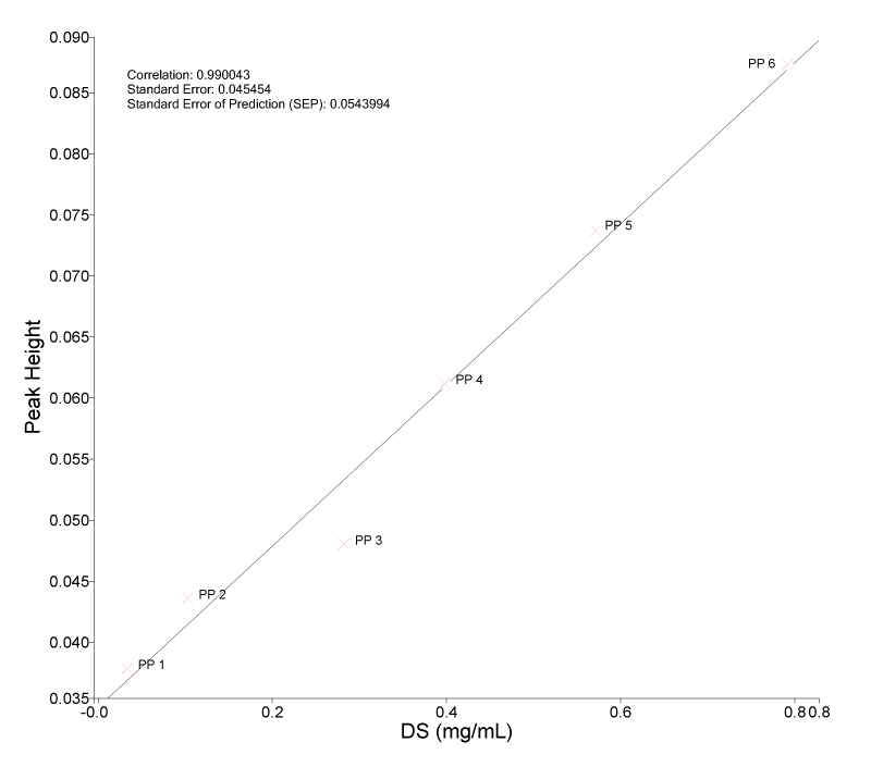
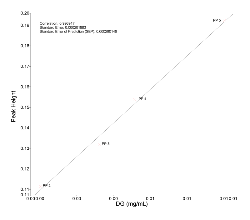
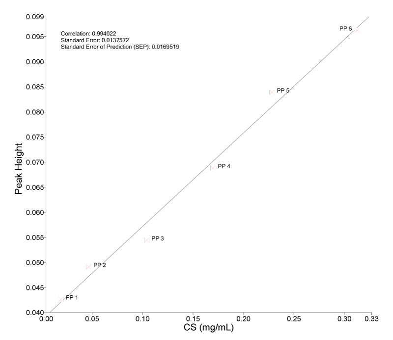
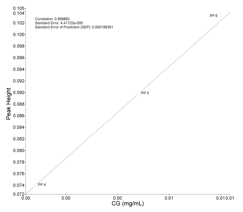
The similar amount in mg of samples used for HPLC were used to make for the spectrum. When this was done, results of HPLC were paired with the spectrum. The standard curve was set by using algorithm Beer’s Law with height for a chosen peak and two base peaks particular for each anthocyanin. The spectrum and the result of HPLC were built up for the correlation in the software “Quant” under function icon ”Review”.
Prediction of anthocyanin content of both sample: Both samples FT 11-15A and FT11-15B from Sabak Bernam were ground into powder form and processed for spectrum. These prepared spectra were imported into the system as independent validation and prediction of the anthocyanin was analysed based on the standard curve (Tables 2,3]. In practice, the spectrum of sample needs to use the similar material which was used for standard curve. Anthocyanin content of the sample was predicted under the system with prediction error. The figures were converted to percentage accordingly.
| Table 2: The standard curve for types of anthocyanins in Raw material. | |||||||
| Delphinidin-3-O-sambubioside | Water extract+ TFA | ||||||
Sample concentration |
FT34/14 | FT10/15 | FT15/16 | FT35/14 | Herbagus | average | sd |
| 1mg/mL | n.a. | 0.0194 | 0.026 | 0.0425 | 0.0031 | 0.02275 | 0.016309 |
| 5mg/mL | 0.1014 | 0.2819 | 0.1648 | 0.1175 | 0.0836 | 0.14984 | 0.079755 |
| 10mg/mL | 0.1155 | 0.2679 | 0.1819 | 0.2316 | 0.1699 | 0.19336 | 0.058657 |
| 15mg/mL | 0.1884 | 0.4264 | 0.2273 | 0.414 | 0.292 | 0.30962 | 0.107604 |
| 20mg/mL | 0.3061 | 0.2832 | 0.4496 | 0.238 | 0.3966 | 0.3347 | 0.086393 |
| 25mg/mL | 0.5401 | 0.925 | 0.5392 | 0.3818 | 1.0903 | 0.69528 | 0.298129 |
| Table 3: the standard curve for types of anthocyanins in water extract+TFA | |
Download Image |
Name DS Type of Fit Linear Calculation Type Peak Height Peak 1 X 1068.38 Peak 1 Base 1 X 1085.69 Peak 1 Base 2 X 978.79 Correlation 0.990043 Standard Error 0.045454 Standard Error of Prediction (SEP) 0.0543994 |
Download Image |
Name DG Type of Fit Linear Calculation Type Peak Height Peak 1 X 1631.12 Peak 1 Base 1 X 1695.30 Peak 1 Base 2 X 1562.53 Correlation 0.996917 Standard Error 0.000201883 Standard Error of Prediction (SEP) 0.000290146 |
Download Image |
Name CS Type of Fit Linear Calculation Type Peak Height Peak 1 X 1062.31 Peak 1 Base 1 X 1085.66 Peak 1 Base 2 X 984.63 Correlation 0.994022 Standard Error 0.0137572 Standard Error of Prediction (SEP) 0.0169519 |
Download Image |
Name CG Type of Fit Linear Calculation Type Peak Height Peak 1 X 1062.69 Peak 1 Base 1 X 1085.66 Peak 1 Base 2 X 975.83 Correlation 0.999883 Standard Error 4.41722e-005 Standard Error of Prediction (SEP) 0.000199391 |
HPLC methodology
Qualitative and quantitative determinations of major constituents of roselle extract was performed on a Waters 2695 Alliance HPLC system®(Waters®, MA, USA) equipped with 996 photodiode array detector and connected to a computer running a Waters Empower 2®software. A C-18 column (Phenomenex, Luna 3_m, 100×4.6mm, i.d.) guarded by a C-18 security guard cartridges (4×3.0mm, i.d.) maintained at room temperature were used. Standards and samples were separated by a mobile phase made of solvent A containing water:TFA (20:0.001; v/v) and solvent B: ACN:TFA (20:0.001; v/v). The following gradient elution was used: 0–2 min, 30% B; 2–10 min, 30–50% B; 10–20 min, 50–95% B and finally washing the column with 95% B for 2 min and reconditioning the column with 30% B isocratic for 2 min. The flow rate was 0.5 ml/min and injection volume for the sample and standards was 10 µl. The peaks were detected at 340 nm.
Five mg of H. sabdariffa dried aqueous extract powder was added into 1 mL of MiliQ water and sonicated for 30 minutes at room temperature. Then the solution was filtered (0.45 μm Nylon) for HPLC analysis.
Authentication
Plant sample labeled FT11-15A was authenticated as Hibiscus sabdariffa var UMKL-1. Plant sample labeled FT11-15B was authenticated as Hibiscus sabdariffa var sabdariffa.
FTIR spectra of H. sabdariffa
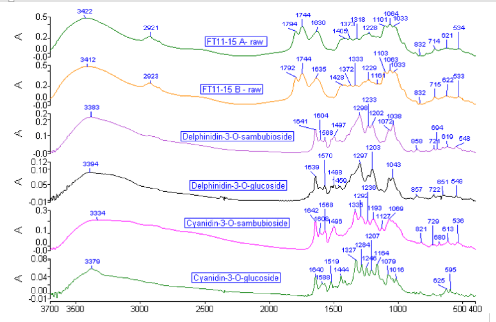
Raw material after decoction: Both varieties of H. sabdariffa raw material after decoction showed a different pattern of spectrum compared with spectra of standard anthocyanins (Figure 1). The basic pattern of H. sabdariffa raw material after decoction spectrum illustrated four sections of wavenumber ranges. The first section started from 3800-2500 cm-1 was normally found in most of the natural products with O-H elongation vibration group (3422 cm-1 at FT11-15A and 3412 cm-1 at FT11-15B) and CH2 stretching band (around 2921 cm-1). The major differentiation of the two varieties of H. sabdariffa began from the second section which was located at the range of 1850-1500 cm-1 with three single peaks of different height. FT 11-15A showed a peak of 1794 cm-1 and indicated the calculated frequencies of parent molecule uracil with assignment δv (C=O), the same peak assignment (1792 cm-1) for FT 11-15 B. Both varieties of H. sabdariffa sample showed peak at 1744 cm-1 referring to stretching of C=O also but more related with ester molecules, whereby the amide I peak of FT11-15A (1630 cm-1) was 5 cm-1 less than FT11-15B. The anthocyanins standards’ main characteristic peaks started from the amide I. Therefore two peaks in front of amide I peak in both samples were not related to anthocyanins. The four anthocyanins standards covered the region 1650-400 cm-1. For identification, there are only 1 to 2 peaks of raw material after decoction peak matching exactly with spectra of the existence of anthocyanin as one of the important contents in H. sabdariffa. In fact, the raw material after decoction contained mixture of multi-compounds. The overlapping of each bond stretching caused the average of peak appeared board and masked many stretching below. Most of the primary metabolites of fatty acid group were found in the range of 1500-1150 cm-1. In this case, the peak of 1404 cm-1 of FT11-15A was 23 cm-1 less compared to FT11-15B. The other different peak was found at 1317 cm-1 of FT11-15A which was 16 cm-1 less than FT11-15B. The saccharide section which started from 1150-400 cm-1 indicated similar peak pattern and position in both samples.
Figure 1: Showed the spectra of FTIR of FT11-15A and FT11-15B raw materials after decoction and compared with 4 types of anthocyanin standards in the range of 4000-400 cm-1.
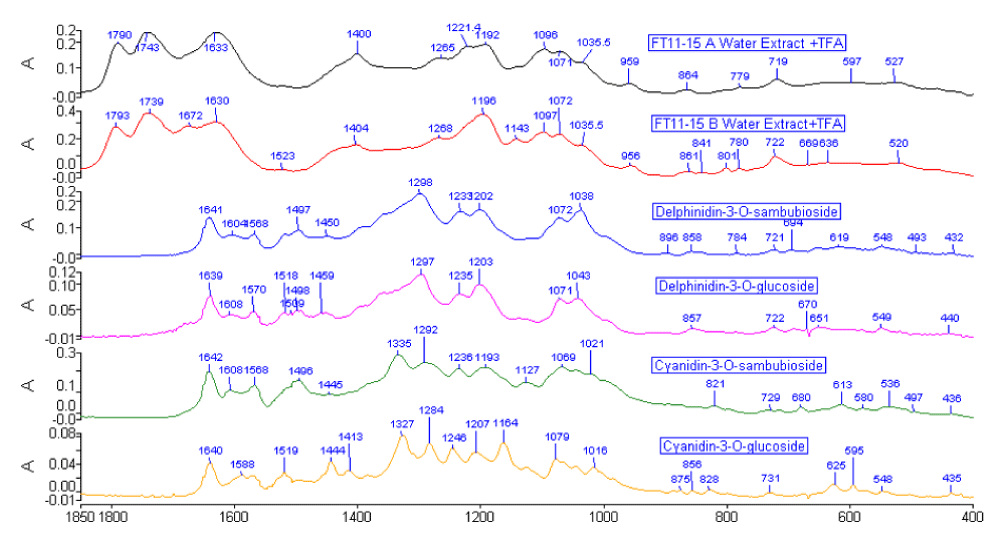
Aqueous extract + TFA: Surprisingly there were many differences between aqueous extract of both samples with 1% TFA (Figure 2) compared to the only aqueous extract (spectrum not shown). One percent of TFA was used in the study for the purpose of stabilising the anthocyanin contents. The appearance of peak height in the region of 1500-1000cm-1 indicated most of the polar compounds have been absorbed in the water and that some proteins were still maintained in the extract. The FTIR aqueous extract + TFA spectrum showed that FT11-15B had a small peak at 1672 cm-1 that did not appear in FT11-15A. Normally, FT11-15B distributed double peak at that region, while FT11-15A showed a single peak with the same height of 1633 cm-1. Eventually the region indicated amide I. Therefore, the peak at 1672 cm-1 was actually derived from the same peak at 1630 cm-1, implying that the protein condition of FT11-15A was more stable than FT11-15B in water extraction. A single sharp peak at 1196 cm-1 of FT11-15B indicated the vibration group of in-plane δ C-H was not affected by water extraction compared to FT11-15A, which derived to a short double peak at 1221 cm-1 and 1192 cm-1. The extra peak at 1143 cm-1 in FT11-15B aqueous extract+TFA spectrum confirmed the different stability condition of flavonoid content mainly anthocyanins in the two varieties of H. sabdariffa. However, both varieties showed similar polysaccharides profiles (peak 959 cm-1 and 956 cm-1) and the maintenance of these chemical constitutes in water extraction. Both sample spectra showed few peaks matched with delphinidin-3-O-sambubioside and delphinidin-3-O-glucoside spectra. The peaks included 1072 cm-1,1038 cm-1 and 721 cm-1. In fact, these three peaks did not appear at raw material spectra. This explained the capability of water extraction enhancing the content of anthocyanins extracted from the raw H. sabdariffa. However, the peak of 1035 cm-1 of both aqueous extract samples spectra showed lower level than 1072 cm-1, opposed to both delphinidin standards. The pattern of both sample spectra from 1192 cm-1 to 1071 cm-1 was very similar to spectrum of cyanidin-3-O-sambubioside, except that the obvious peak 1096 cm-1 was absent from this anthocyanin standard. There was only a peak of 1079 cm-1 of cyanidin-3-O-glucoside that closely related to the samples. Comparative spectra were therefore useful in determining the existence of anthocyanins content in both H. sabdariffa samples.
Figure 2: Showed the FTIR spectra of FT11-15A and FT11-15B aqueous extract +TFA compared with 4 types of anthocyanin standards in the range of 1850-400 cm-1.
2DIR synchronous and asynchronous spectrum
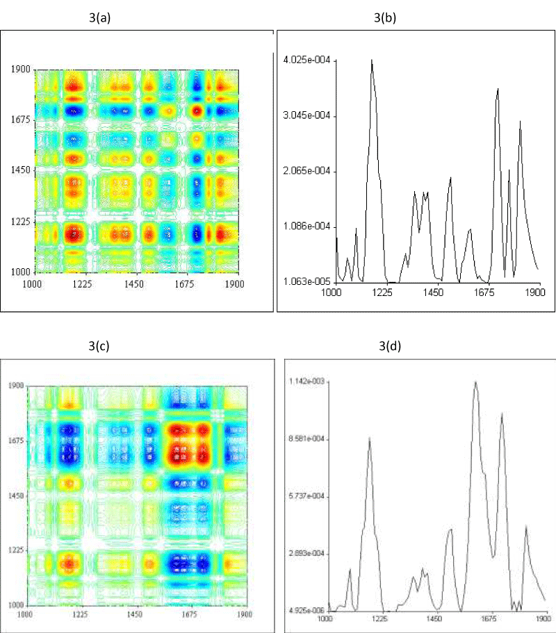
Raw material after decoction: Figure 3a-d) provides a holistic chemical profile of the raw material after decoction. The overview of the configuration before the extraction enhanced the reality of macro-fingerprint by way of molecular structure. The FT11-15A synchronous 2DIR raw material after decoction spectrum presented more red spots than FT11-15B in the range of 1900-1000cm-1, and they were scattered in almost a quarter of the 2D spectrum. This implicated that at least 70% of the components in FT11-15A reacted actively during the thermal perturbation and many overlapping peaks were found. For comparison, the synchronous 2DIR spectrum of FT11-15B (Figure 3c), a small area of red spots located at the top right corner was analysed. Detailed interpretation indicated FT11-15A had 8 auto peaks instead of 5 auto peaks in FT11-15B. In addition, FT11-15A created 15 positive cross peaks and 12 negative cross peaks. The important correlation squares were built among auto peak 1814 cm-1, cross peak (1716,1814), auto peak 1716 cm-1, cross peak (1814,1716); auto peak 1814 cm-1, cross peak (1504,1814), auto peak 1504 cm-1, cross peak ( 1814,1504); auto peak 1504 cm-1, cross peak (1400, 1504), auto peak 1400 cm-1, auto peak (1504,1400); auto peak 1504 cm-1, cross peak ( 1169, 1504), auto peak 1169cm-1, cross peak ( 1504,1169) and respective biggest square was auto peak 1814 cm-1, cross peak (1169, 1814), auto peak 1169 cm-1, cross peak (1814, 1169) . The correlation between the area of peak 1814 cm-1, 1716 cm-1, 1504 cm-1 and 1169 cm-1 represented the vibration occurring concurrently with the functional perturbed stretching of carbonyls, vibration of ketone, amide II band and probably in plane C-H bending of phenyl. On the other hand, FT11-15B showed 3 main positive cross peaks and they were separated with correlation squares among auto peak 1716 cm-1, cross peak (1610, 1716), auto peak 1610 cm-1, cross peak (1716,1610); auto peak 1502 cm-1, cross peak (1171, 1502), auto peak 1171 cm-1, cross peak (1502, 1171). The differences of the two varieties in 2DIR in this range was that FT11-15A had more active components that reacted evenly, but FT11-15B showed only few active components around the peak of 1675 cm-1 during the perturbation.
Figure 3: 2DIR Synchronous and auto peak spectra of FT11-15A and FT11-15B raw material in the range of 1900-1000 cm-1.
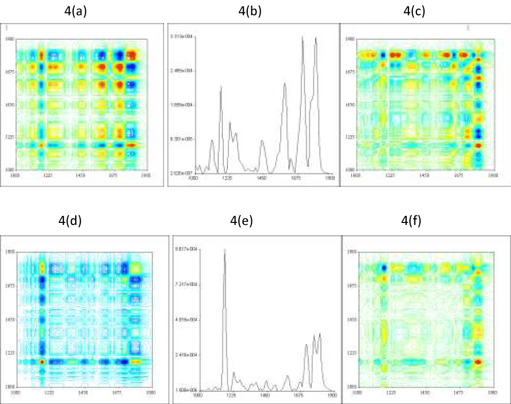
FT11-15A aqueous extract + TFA: Figure 4a shows the 2DIR synchronous spectrum of FT11-15A aqueous extract +TFA in the range of 1900-1000 cm-1. There were 7 auto peaks (Figure 4b) reported along the diagonal line. The correlation square among peak auto peak 1795 cm-1, cross peak (1585,1795), auto peak 1585 cm-1, cross peak (1795, 1585); auto peak 1795 cm-1, cross peak (1171, 1795), auto peak 1171 cm-1, cross peak (1795, 1171); auto peak 1585 cm-1, cross peak (1171, 1585), auto peak 1171 cm-1, cross peak (1585,1171); auto peak 1710 cm-1, cross peak (1592, 1710), auto peak 1592 cm-1, cross peak (1710,1592) were the most active sites with highest intensity of auto peak. The association of correlation square explored a related variation of functional group among the components. The Noda’s law for 2DIR interpretation included synchronous and asynchronous spectrum. For the water extract which had eliminated most of the primary metabolites, the asynchronous spectrum (Figure 4c) was coordinated in correlation. The cross peak at (1169,1795) was positive in synchronous spectrum but negative in asynchronous spectrum, and it could therefore be assumed that the auto peak 1795 cm-1 had the privilege to initial the vibration before auto peak 1169 cm-1.
FT 11-15 B aqueous extract +TFA: There were some differences of 2DIR profile for FT11-15B aqueous extract+TFA. Figure (4d) presented a huge correlation square among auto peak 1814 cm-1 (4e), cross peak (1167, 1814), auto peak 1167 cm-1 and cross peak (1814, 1167). The correlation between each main peak was the negative cross peaks playing the intermediate roles. The asynchronous spectrum (figure 4f) showed the opposite of figure (4e). The clear positive cross peak at (1167, 1814) in figure (4d) had turned negative cross peak in figure (4f). It is important to state that peak of 1814 cm-1 assigned for vibration C=O group begun to vibrate followed by peak of 1167 cm-1 assigned for in-plane δ C-H, characteristic of p-substitution group in water extract during the thermal perturbation.
Figure 4: (4a) showed the 2DIR synchronous spectrum of FT11-15A aqueous extract+ TFA in the range of 1900-1000 cm-1; (4b) its auto peak spectrum and (4c) the 2DIR asynchronous spectrum of FT11-15A aqueous extract+TFA in the same range. (4d) showed the 2DIR synchronous spectrum of FT11-15B aqueous extract +TFA in the range of 1900-1000 cm-1; (4e) its auto peak spectrum and (4f) the 2DIR asynchronous spectrum of FT11-15B aqueous extract +TFA in the same range.
The quantification of anthocyanin content in two groups of H. sabdariffa samples using software “Quant”
Table 4 presented the quantitative result of 4 types of anthocyanin of FT11-15A and FT11-15B based on the established standard curve. In this small-scale analysis, the content of anthocyanin of both H. sabdariffa raw material after decoction sample in descending amount was delphinidin-3-O-sambubioside, cyanidin-3-O-sambubioside, delphinidin-3-O-glucoside and lastly cyanidin-3-O-glucoside. Both samples raw materials after decoction demonstrated small differences in all the anthocyanins amount. FT11-15A reported 0.0113 mg/mL of delphinidin-3-O-sambubioside more than FT11-15B, with almost same % prediction error. FT11-15A also recorded more delphinidin-3-O-glucoside than FT11-15B. On the other hand, FT11-15B contained 0.0043 mg/mL of cyanidin-3-O-sambubioside more than FT11-15A. Cyanidin-3-O-glucoside of both samples showed lower difference and was the lowest among the amount of anthocyanin in H. sabdariffa raw material samples. The result showed that the raw material after decoction has very low amount of anthocyanin detected by spectroscopy method and could be due to the presence of mixture of all ingredients in the raw material. This scenario was obvious when comparing the amount of cyanidin-3-O-sambubioside of raw material after decoction and water extract plus TFA. The content of anthocyanin was mixed with other contents of H. sadbariffa and the spectrum peak selected for the anthocyanin determination have been overlapped with others compound bonding. The extraction by water has eliminated most of the unwanted contents especially the debris, fiber and explored the second metabolism products. Therefore, higher anthocyanin content of water extract was detected than in the raw material after decoction. In fact, aqueous extract+TFA especially from FT11-15B achieved the highest amount of delphinidin-3-O-sambubioside. In addition, the % prediction error of the content of cyanidin-3-O-glucoside of aqueous extract+TFA was 1-5 times more than the raw. This could be due to the extremely small quantities of this type of anthocyanin compared to the other anthocyanins in the sample itself or the factors of pre and post-harvest. It is interesting that in water extract, majority content of anthocyanin in FT11-15B was more than FT11-15A.
| Table 4: Result of the 4 types of anthocyanins of the sample FT11-15A and FT11-15B from Sabak Bernam, Selangor. * indicated the prediction error. %= percentage of prediction error. | ||||||
| Anthocyanin | Type of sample | sample | mg/mL | % difference | * | % prediction error |
| Delphinidin 3-O-sambubioside | Raw material | FT 11-15A | 0.5959 | FT11-15A > FT 11-15B = 1.13% |
0.1503 | 25.2 |
| FT 11-15B | 0.5846 | 0.1495 | 25.5 | |||
| Water extract +TFA | FT 11-15A | 0.5950 | FT 11-15B > FT11-15A = 3.68% |
0.1933 | 32.5 | |
| FT 11-15B | 0.6318 | 0.2052 | 32.5 | |||
| Delphinidin 3-O-glucoside | Raw material | FT 11-15A | 0.0055 | FT 11-15A > FT11-15B = 0.07% |
0.0010 | 18.2 |
| FT 11-15B | 0.0048 | 0.0010 | 20.8 | |||
| Water extract + TFA | FT 11-15A | 0.0050 | FT 11-15B > FT 11-15A = 2.01% |
0.0012 | 24.0 | |
| FT 11-15B | 0.0251 | 0.0047 | 18.7 | |||
| Cyanidin 3-O sambubioside | Raw material | FT 11- 15A | 0.2224 | FT 11-15B > FT 11-15A= 0.43% |
0.0447 | 20.1 |
| FT 11- 15B | 0.2267 | 0.0449 | 19.8 | |||
| Water extract + TFA | FT 11-15A | 0.7447 | FT 11-15B> FT 11-15A= 8.98% |
0.2681 | 36.0 | |
| FT 11-15B | 0.8345 | 0.3020 | 36.2 | |||
| Cyanidin 3-O glucoside | Raw material | FT 11-15A | 0.0044 | FT 11-15B > FT 11-15A = 0.01% |
0.0070 | 15.9 |
| FT 11-15B | 0.0045 | 0.0070 | 15.6 | |||
| Water extract + TFA | FT 11-15A | 0.0046 | FT 11- 15A > FT 11-15B= 0.03% |
0.0036 | 78.3 | |
| FT 11-15B | 0.0043 | 0.0035 | 81.4 | |||
The study successfully compared the two varieties of H. sabdariffa collected from Sabak Bernam, Selangor using the two stages of FTIR and 2DIR correlation spectroscopy and quantified the amount of anthocyanin by combination of HPLC and spectra in “Quant” software. The FTIR spectral reported FT11-15B raw material after decoction contained higher wavenumber from 1500 cm-1 to 1200 cm-1 when compared to the same functional group in FT11-15A. Actually, the FTIR spectrum of FT11-15B raw material after decoction had scored a correlation rate of 0.990173 compared to FT11-15A. Nonetheless, FT11-15A showed the stability of amide I group in its aqueous extract. More distinguished areas of spectrum were noticed in H. sabdariffa aqueous extract due to high anthocyanin aqueous solubility. In 2DIR spectrum, FT11-15A showed the positive auto peak and cross peak scattering in the range of 1900-1000 cm-1 evenly, implying that more active components were involved, compared to FT11-15B especially the anthocyanin content. The application of 2DIR spectrum is the second step to enhance the discrimination of both varieties.
The combination of quantity method typically established a basic of prediction for anthocyanin of other H. sabdariffa samples from different locations. As the first run with HPLC, the accuracy of the quantity was initially developed. Spectra of the sample either in the medium of raw material or extract were compatible to the results of HPLC accordingly. The software that combined the advantages of both different techniques and produced the detail analysis of anthocyanins determination. The established sample H. sabdariffa standard curve can be used for other location anthocyanin sample prediction. This method has expediated analysis, as the sample for anthocyanin prediction has excluded a step of chromatography. This is a solution that can be used for large scale analysis of samples, cost-effective and time saving.
Both sample groups were successfully discriminated using the combination of chromatography and two steps of spectroscopy methods, indicating that both sample groups contained different ingredients even though they are from similar location. There could be possibility that the factor of two different varieties and the pre and post-harvest of condition in sample processing. The quantification study concluded that both samples showed highest amount of delphinidin-3-O sambubioside, followed by cyanidin-3-O-sambubioside, delphinidin-3-O-glucoside and cyanidin-3-O-glucoside. The anthocyanin content of FT11-15A and FT11-15B varied with the types of anthocyanins analyzed using the same method.
We would like to thank the Director General of Health, Malaysia for permission to publish this paper, and the Director of Institute for Medical Research (IMR), Kuala Lumpur for support of this project. This work was financially supported by the NKEA AGRICULTURE (EPP#1), NKEA Research Grant Scheme (NRGS) (Grant No: NH1014D060).
- Mariod AA, Saeed Mirghani ME, Hussein I, Mariod AA, Saeed Mirghani ME, et al. Hibiscus sabdariffa L. Roselle. Unconventional Oilseeds and Oil Sources. 2017; 59-65.
- Riaz G, Chopra RA. review on phytochemistry and therapeutic uses of Hibiscus sabdariffa L. Biomedicine & Pharmacotherapy, 2018; 102: 575-586. PubMed: https://www.ncbi.nlm.nih.gov/pubmed/29597091
- Fallahi HR, Ramazani SHR, Ghorbany M, Aghhavani-Shajari, M. Path and factor analysis of roselle (Hibiscus sabdariffa L.) performance. Journal of Applied Research on Medicinal and Aromatic Plants. 2017; 6: 119-125.
- Cercato LM, White PAS, Nampo FK, Santos MRV, Camargo EA. A systematic review of medicinal plants used for weight loss in Brazil: Is there potential for obesity treatment? Journal of Ethnopharmacology. 2015; 176, 286-296. PubMed: https://www.ncbi.nlm.nih.gov/pubmed/26520790
- Hirunpanich V, Utaipat A, Morales NP, Bunyapraphatsara N, Sato H,et al. Hypocholesterolemic and antioxidant effects of aqueous extracts from the dried calyx of Hibiscus sabdariffa L. in hypercholesterolemic rats. Journal of Ethnopharmacology. 2006; 103, 252-260. PubMed: https://www.ncbi.nlm.nih.gov/pubmed/16213683
- Ojeda D, Jiménez-Ferrer E, Zamilpa A, Herrera-Arellano A, Tortoriello J, et al. Inhibition of angiotensin convertin enzyme (ACE) activity by the anthocyanins delphinidin- and cyanidin-3-O-sambubiosides from Hibiscus sabdariffa. Journal of Ethnopharmacology. 2010; 127: 7-10. PubMed: https://www.ncbi.nlm.nih.gov/pubmed/19808084
- Yang B, Liu H, Yang J, Gupta VK, Jiang Y. New insights on bioactivities and biosynthesis of flavonoid glycosides. Trends in Food Science & Technology. 2018; 79: 116-124.
- Zheoat AM, Gray AI, Igoli JO, Ferro VA, Drummond RM. Hibiscus acid from Hibiscus sabdariffa (Malvaceae) has a vasorelaxant effect on the rat aorta. Fitoterapia. 2019; 134: 5-13. PubMed: https://www.ncbi.nlm.nih.gov/pubmed/30690125
- Jackson RD, Isidore B, Cates RL. Are plant-soil dynamics different in pastures under organic management? A review. Agriculture, Ecosystems & Environment. 2019; 279: 53-57.
- Juhari NH, Bredie WLP, Toldam-Andersen TB, Petersen MA. Characterization of Roselle calyx from different geographical origins. Food Research International. 2018; 112, 378-389. PubMed: https://www.ncbi.nlm.nih.gov/pubmed/30131149
- Sharma HK, Sarkar M, Choudhary SB, Kumar AA, Maruthi RT, et al. Diversity analysis based on agro-morphological traits and microsatellite based markers in global germplasm collections of roselle (Hibiscus sabdariffa L.). Industrial Crops and Products. 2016; 89: 303-315.
- Osman Mohamad, Saberi S, Nezhadahmadi Arash, Faruq Golam. Development and Evaluation of Fruit Related Morphological and Physico-Chemical Characteristics in Three Roselle Mutants. Pakistan Journal of Nutrition. 2014; 13.
- Ma’arup R, Aziz MA, Osman M. Development of a procedure for production of haploid plants through microspore culture of roselle (Hibiscus sabdariffa L.). Scientia Horticulturae. 2012; 145: 52-61.
- Mahadevan N, Shivali A. and Kamboj P. Hibiscus sabdariffa Linn.-An overview. Natural Product Radiance. 2009; 8: 77-83. https://essentia.com.br/images/artigos/ativos-nov-2016/Tintura%20Hisbiscus.pdf
- Tham TC, Ng MX, Gan SH, Chua LS, Aziz R, et al. Impacts of different drying strategies on drying characteristics, the retention of bio-active ingredient and colour changes of dried Roselle. Chinese Journal of Chemical Engineering. 2018; 26: 303-316.
- Lu, Y., Wu, N., Fang, Y., Shaheen, N., & Wei, Y. An automatic on-line 2,2-diphenyl-1-picrylhydrazyl-high performance liquid chromatography method for high-throughput screening of antioxidants from natural products. Journal of Chromatography A. 2017; 1521: 100-109. PubMed: https://www.ncbi.nlm.nih.gov/pubmed/28939230
- Jiao L, Guo Y, Chen J, Zhao X, Dong D. Detecting volatile compounds in food by open-path Fourier-transform infrared spectroscopy. Food Research International. 2019; 119: 968-973. PubMed: https://www.ncbi.nlm.nih.gov/pubmed/30884737
- Smith MJ, Holmes-Smith AS, Lennard F. Development of non-destructive methodology using ATR-FTIR with PCA to differentiate between historical Pacific barkcloth. Journal of Cultural Heritage. 2019.
- Hua R, Sun SQ, ZhouQ, Noda I, Wang BQ. Discrimination of Fritillary according to geographical origin with Fourier transform infrared spectroscopy and two-dimensional correlation IR spectroscopy. Journal of Pharmaceutical and Biomedical Analysis. 2003; 33: 199-209. PubMed: https://www.ncbi.nlm.nih.gov/pubmed/12972085
- Zuo L, Sun S, Zhou Q, Tao J, Noda I. 2D-IR correlation analysis of deteriorative process of traditional Chinese medicine ‘Qing Kai Ling’ injection. Journal of Pharmaceutical and Biomedical Analysis. 2003; 30: 1491-1498. PubMed: https://www.ncbi.nlm.nih.gov/pubmed/12467920
